Register for this event
Please complete all sections below
Sorry! Registration is completed!
The school has been oversubscribed so unfortunately we are forced to stop registration of new participants.
The school starts in:
-
00
days
-
00
hours
-
00
minutes
-
00
seconds
What is this school and for whom?
The international school "Modern cryoelectron microscopy" is organized by the Moscow Institute of Physics and Technology and the Lomonosov Moscow State University. The purpose of the event is to familiarize students, graduate students, researchers of academic and scientific organizations with a modern and dynamically developing structural research method - cryoelectron microscopy. The school is a three-day intensive course on the methods used in cryoelectron microscopy, including theoretical training, an overview of the modern world instrumentation infrastructure, sample preparation methods, conducting experiments and analyzing the results. The spectrum of topics covered suits attendees that are new to the field as well as those for whom the technique is already of growing importance in their research and who wish to acquire an in-depth understanding of the methodology. Educational lectures and master classes will be given. Leading researchers in the field of cryoelectron microscopy from Russia, Great Britain, Switzerland, Germany and France will be tutors of the school.
The number of participants is limited. School participants are expected to give a presentation of their current project.
The language of the school is English.
Topics
The advantages of modern cryo-electron microscopes
Why is this topic so popular in recent years?
The heterogeneity of the samples, the problems associated with biological samples
Tomography and single particle analysis
Validation of structures, how to evaluate their correctness?
What determines method resolution?
Tutors
All invited tutors is experienced lecturers and instructors are all users of cryoelectron microscopy infrastructure and methods.
Alexander Vasiliev
National Research Center "Kurchatov Institute" / Russia
Elena Orlova
Birkbeck University of London / United Kingdom
Ambroise Desfosses
Institut de Biologie Structurale / France
Evgeniya Pechnikova
ThermoFisher Scientific / United Kingdom
Agustin Avila-Sakar
Gatan, Inc. / USA
Frédéric Leroux
Leica Microsystems / Belgium
Olga Sokolova
Moscow State University / Russia
Vadim Cherezov
Bridge Institute, USC, USA / Moscow Institute of Physics and Technology, Russia
Grigory Sharov
MRC Cambridge / United Kingdom
Gunnar Schröder
Forschungszentrum Jülich / Germany
Albert Guskov
University of Groningen / Netherlands
Committees
ORGANIZING COMMITTEE
- Dr. Andrey Rogachev
- MIPT / JINR - Co-chairman
- Dr. Valentin Borshchevskiy
- MIPT - Co-chairman
- Natalia Iakovleva
- MIPT – Scientific secretary
- Dr. Alexey Mishin
- MIPT
- Dr. Ivan Gushchin
- MIPT
- Dr. Maxim Nikitin
- MIPT
- Ivan Okhrimenko
- MIPT
- Eugenia Chirkina
- MIPT
- Ekaterina Donskaya
- MIPT
- Dr. Alina Remeeva
- MIPT
PROGRAM COMMITTEE
- Prof. Dr. Valentin Gordeliy
- IBS, France / Forschungszentrum Juelich, Germany – Co-chairman
- Prof. Dr. Olga Sokolova
- Lomonosov Moscow State University, Russia – Co-chairman
- Prof. Dr. Elena Orlova
- University of London, UK
- Prof. Dr. Vadim Cherezov
- USC, USA / MIPT, Russia
- Prof. Dr. Marat Yusupov
- Kazan Federal University, Russia
- Prof. Dr. Carsten Sachse
- Forschungszentrum Jülich, Germany
Important Dates
15 February
First announcement
20 February
Open registration
11 March
Second Information Bulletin
18 March
The school has been oversubscribed so unfortunately we are forced to stop registration of new participants
31 March
Visa Deadline
20 May
Registration deadline
30 May - 01 June
Working days of the international school
SCHEDULES
30th May 2019
TOPICS:
The history of the method
Why is this topic so popular in recent years?
Examples of use for biological research
Theoretical background
Anatomy of cryoelectron microscopes
-
10:35 - 12:05
90 min
Olga Sokolova
Moscow State University / Russia
Cryo-EM: from the XX century into the XXI
In the 60-s of the XX century, the background for the current rapid development of cryo-electron microscopy has been established. At that time, single particle EM was used to determine 3D structures of large molecules in negative contrast. Later, cryo-EM helped to obtain three-dimensional structures of molecules with higher resolution. In the XXI century, accelerating voltages of electron microscopes have increased, field emission cathodes and modern electron-optical systems have appeared, improving the characteristics of the electron beam. Direct electron detectors opened up a new way of collecting the images. By 2019, more than 8000 protein 3D structures have been sub-mitted to EMDB, including de-novo determined structures. Due to the constant modifi-cation of computer software, fully automated data collection and processing cryo-EM are now accessible to the wider scientific community.
-
12:25 - 13:55
90 min
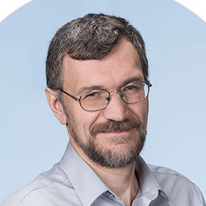
Vadim Cherezov
Bridge Institute, USC, USA / Moscow Institute of Physics and Technology, Russia
Structural Studies of G Protein-Coupled Receptors by Cryo-EM
G Protein-Coupled Receptors (GPCRs) are cellular gatekeepers that regulate a variety of physiological processes in the human body and serve as attractive pharmaceutical drug targets. Structure-function studies of this superfamily have been enabled in 2007 by multiple breakthroughs in technology that included receptor stabilization, crystallization in a membrane environment, and microcrystallography. Recent resolution revolution in Cryo-EM further accelerated structural studies of GPCR superfamily, in particular structure determination of signaling complexes of receptors with their cognate G proteins. This talk will summarize recent Cryo-EM work on GPCRs, highlighting important advancements in G protein engineering, development of antibodies that stabilize the heterotrimeric G proteins, and selection of appropriate solubilization strategies. These structures shed light on the mechanism of signal transduction and G protein selictivity and provide important clues for designing biased ligands.
-
15:00 - 16:30
90 min
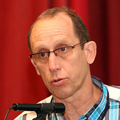
Alexander Vasiliev
National Research Center "Kurchatov Institute" / Russia
New methods of electron microscopy of functional materials
During last years, the electron microscopy (EM) instrumentation and techniques have changed drastically. Spherical aberration correctors (Cs-correctors) of electromagnetic lenses, electron gun monochromators, new high-performance X-ray detectors improved the special resolution, speed and sensitivity of EM. Electron-optical systems make it possible to focus the beam on the sample with the probe diameter less than 1 Å, and the ultra-pure vacuum al-lows obtaining nanodiffraction patterns from particles of subnanometer size. The Cs correct-ed condenser systems permit to obtain electron-beam images with a high resolution in Z-contrast mode with a sub angstrom resolution. The efficiency of large area energy dispersive X-ray (EDX) silicon drift detectors is up to 106 pulses per second, which is 2 orders higher than that of single crystal Si (Li) detectors keeping the same energy resolution. With these detectors now is possible to obtain distribution maps of elements with atomic resolution. The use of new methods for sample preparation for transmission microscopy using a focused ion beam (FIB) in dual-beam electron-ion microscopes changed the accuracy of selecting the area for thin section close to several nm, which is often necessary in the study of multiphase mate-rials. Dual-beam Ems gave rise to the “slice-and-view” method, which allows one to reconstruct three-dimensional microstructural materials and, upon the use of EDX detectors to restore in 3D the phase composition. The next group of new methods is the use of cryo-EM. Specimens for cryo-EM are fast freezed in amorphous ice to liquid nitrogen temperature. That low down the radiation damag-es and stability the material under the e--beam. A number of examples of studies using these methods will be presented.
-
17:00 - 18:30
90 min
Evgeniya Pechnikova
ThermoFisher Scientific / United Kingdom
Basics of transmission electron microscopy: organization of microscope and image formation
Transmission electron microscopy (TEM) is a powerful technique that finds its implementation in different fields of research.
Architecture of transmission electron microscope includes following parts: electron source, column (condenser system, objective lens, projection system) , energy filter (optionally), detector. Performance of a microscope depends on all of these parts.
During the lecture we will go into details how each part of a microscope works, and how they contribute to image formation.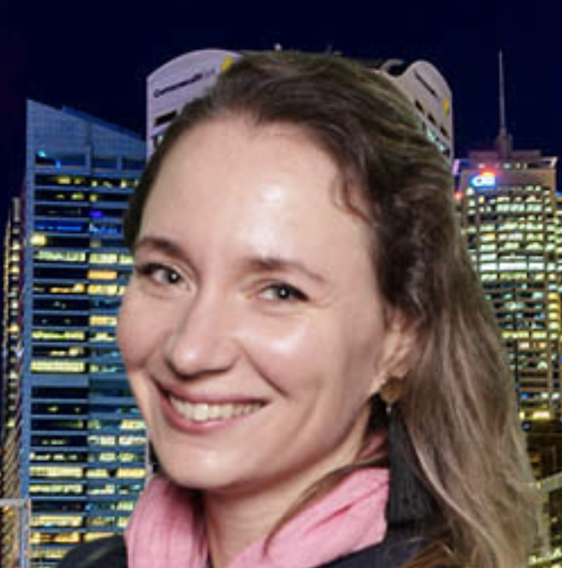
31th May 2019
TOPICS:
Sample preparation
Features and various methods of experiments
Tomography and single particle analysis
The heterogeneity of the samples, the problems associated with biological samples
-
10:00 - 13:20
200 min
Frédéric Leroux
Leica Microsystems / Belgium
PART 1. Conventional to cryo preservation
PART 2. Cryo processing (cemovis, freeze substitution for 3D, freeze fracturing, cryo FIB)
PART 3. CLEM & emerging technologiesConventional electron microscopy relies on chemical fixation of biological samples. Such processing steps inevitably influence the observed ultrastructure. Although still relevant today, an alternative workflow to capture the native state of biological samples becomes increasingly important. During the lectures a stepswise approach will be used to explain the available specimen preparation workflows and how they are relevant in solving the most challenging biological questions. Conventional chemical fixation will be the starting point and the lectures will cover following topics: sample preservation, chemical and physical; sample processing under room and cryogenic conditions (sectioning, fracturing, ion beam milling), sample screening (correlative microscopy).
(Include coffee brake)
-
14:20 - 16:20
120 min
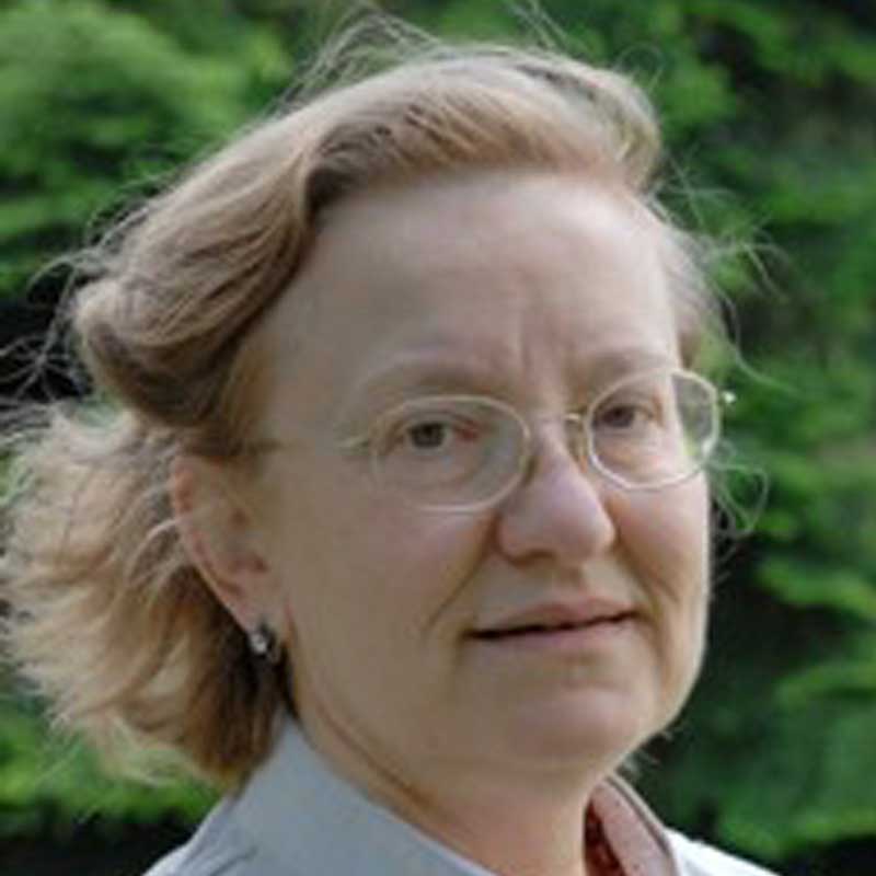
Elena Orlova
Birkbeck University of London / United Kingdom
Revelation of function of bio-complexes through analysis of their dynamics by cryo-electron microscopy and image processing
In living organisms, biological macromolecules are intrinsically flexible and naturally exist in multiple conformations. Cryo electron microscopy, visualises bio-complexes in nearly native conditions. The advances made during the last decade in electronic technology and software development allowed us to identify structural variations in complexes at nearly atomic resolution. EM studies based on single-particle methods (SPA) developed several approaches for the separation of different conformational states and therefore disclosure of the mechanisms for functioning of complexes. To resolve different states of molecules requires the examination of large datasets, sophisticated programs and significant computing power. Some methods are based on analysis of two-dimensional images; while others are based on three-dimensional studies. Here, will be given an overview of basic principles implemented in the various techniques that are currently used in the analysis of structural conformations and provide some examples of successful applications of these methods in structural studies of biologically significant complexes.
-
16:50 - 18:50
120 min
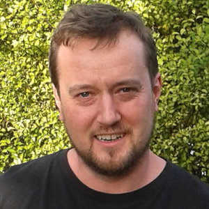
Ambroise Desfosses
Institut de Biologie Structurale / France
Tomography and single-particle reconstruction : basics, recent developments and future directions
This lecture will cover the basic principles of electron tomography and single-particle reconstruction, with examples of applications. Strategies to process the movies generated by direct electron detectors will be discussed, in particular regarding motion correction and dose weighting. Adaptation of the workflow for imaging/processing small proteins and large complexes will be detailed. A particular emphasis will be given to recent developments, current limitations and future directions of cryo-EM for solving biological structures.
1st June 2019
TOPICS:
Resolution
Detectors
Working with images
Programs and algorithms
-
10:00 - 11:30
90 min
Agustin Avila-Sakar
Gatan, Inc. / USA
Automated Recording of Single Particle Cryo-EM Data
Cryo-EM is the low-dose imaging technique par excellence. The very high sensitivity of biological macromolecules to accelerated electrons imposes a limitation on the exposure that can be used to record data directly from them. As a consequence, electron images of biological samples are inherently noisy. Since its early beginnings, cryo-EM had to cope with the challenge of taking pictures of almost invisible objects without even being able to attempt seeing them first in any detail (to minimize radiation damage), and to combine data from a very large number of noisy images (to harvest the signal from within the noisy data). Throughout its existence, Cryo-EM has benefited enormously from advances in electron optical instrumentation, computer power and the development of new technologies. Most recently, the advent of CMOS* direct electron detection propelled cryo-EM into an astonishingly fast maturation phase, often referred to as the “Resolution Revolution”, which brought the technique to the very forefront of structural biology. Such a leap, in turn, has created the need for higher standards of automation and data throughput. This lecture will explain the rationale behind the methods of data recording in single particle cryo-EM, and how they are shaping automated data acquisition schemes. The steps involved in the setup and execution of fully automated cryo-EM sessions will be described and illustrated using Gatan’s single particle data acquisition software Latitude-S. *CMOS = complementary metal oxide semiconductor
-
12:00 - 13:50
110 min
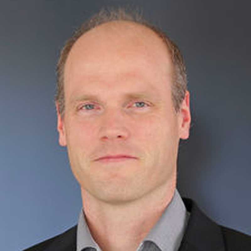
Gunnar Schröder
Forschungszentrum Jülich / Germany
PART 1.
Helical Reconstructions and Atomic Model Building and RefinementIn this lecture, helical reconstructions will be introduced, from particle picking to reconstructions, including some applications to amyloid fibrils. Later map sharpening procedures will be discussed, in particular the VISDEM sharpening method. The model building software emfasa will be presented. Furthermore, a cross-validation method is resented to prevent overfitting during structure refinement.
-
16:10 - 17:10
60 min
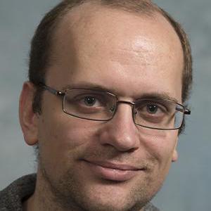
Grigory Sharov
MRC Cambridge / United Kingdom
PART 1 "Getting the best data from your scope"
Choice of microscope and detector for screening or data collection. How does it depends on specimen? How do I choose magnification and electron dose? How to get best images from your TEM? A brief overview of how microscope, detector, sample, specimen, and image processing parameters and decisions might affect your resolution.
-
18:30 - 20:00
90 min
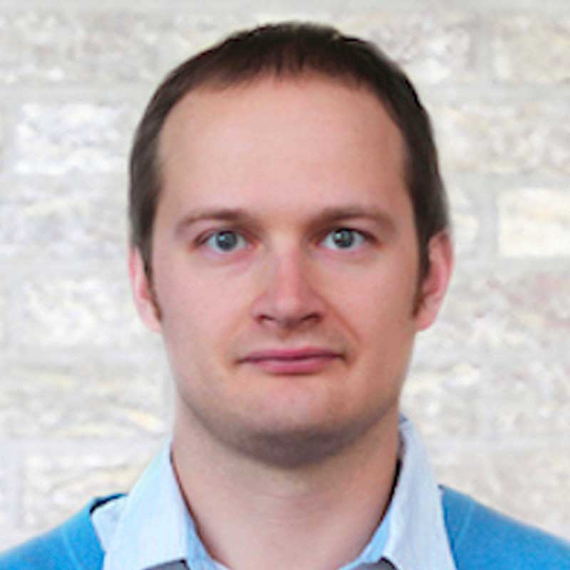
Albert Guskov
University of Groningen / Netherlands
Electron Microscopy of Biomembranes and Their Components
After the reconstruction, users end up with electron density maps. This is where the interpretation of data begins, thus one has to be extremely careful and, in a way, responsible to construct as accurate model as possible. In this lecture the students will learn about the good practices of model building at different levels of resolution (high, moderate, low), as well as about refinement both in real and reciprocal space. Ultimately the overview of current progress in validation of generated models will be given.
Registration Fees
Full registrants are welcome at general activities, have access to the lecture halls, will be provided with refreshments at two coffee breaks each day and Endless opportunities to network with colleagues from around the globe. The Conference Dinner or Lunch is NOT included in any registration types but you will have time to “lunch break” in the program. Also the conference fee does not include travel and accommodation.
At the moment, the organizers are deciding on the possibility of accommodating participants on the MIPT campus. This feature and cost will be announced later.
Cost of participation as a listener:
Before 31 March 2019 — 3 000 rubles
After 31 March 2019 — 6 000 rubles
You can pre-register now and pay the registration fee later.
* Information for MSU and MIPT students. For details on the terms of participation please contact to the organizing committee.
Venue
Moscow Institute of Physics and Technology (“Phystech”) is the leading institute of higher education in Russian Federation training highly-qualified specialists in various areas of modern science and technology. The institute has a rich history. Its founders and early staff members were Nobel Prize winners P.L Kapitsa, N.N Semenov, and L.D. Landau. Nobel Prize winners are among its graduates as well. Many “Phystech” professors are leading scientists in Russia, among which are more than 80 academics and corresponding members of the Russian Academy of Sciences. From the beginning, Moscow Institute of Physics and Technology has used a unique system for training specialists, known commonly as the “Phystech System,” which combines fundamental education, engineering disciplines, and student scientific research. Now the Institute is developing unique scientific infrastructure to carry out the cutting edge research in many fields of science.
Contact Us
Moscow Institute of Physics and Technology (State University)
9 Institutskiy per.,Dolgoprudny, Moscow Region, 141700, Russian Federation
school@cryo-em.ru
We start May,30th at 9.00 am, “Phystech.Arctica” bld. (“Физтех.Арктика”)
(Moscow region, Dolgoprudny city, Nauchny lane, 4)


